There’s something remarkable about seeing a tiny baby’s heart beat during an ultrasound scan for the very first time. While it pounds away at an impressive pace, the child growing within us becomes more tangible. And now, thanks to the marvels of technology and some very dedicated doctors, we can see these little human beings in a whole new light.
A team of researchers at King’s College London and Guy’s and St Thomas’s in London have been working on building more detailed pictures of the baby’s heart while in utero to help provide better treatment for those born with congenital heart disease. Although ultrasounds have previously given doctors a lot of insight into the unborn baby’s health, their tiny hearts beat so quickly it’s tricky to detect any issues the child might have later on in life. So in the researchers’ study, pregnant women were given an MRI scan that provided a number of 2D images of the baby’s heart, which, coupled with some impressive computer software, enabled doctors to build up a 3D-model replica of the heart.
One life-saving case was of baby Violet-Vienna, which was reported in the BBC. During the regular 20-week scan, doctors could detect some issues with the blood vessels that went around the baby’s heart, narrowing her aorta. Her mom, Kirbi-Lea Pettitt, joined the study and doctors were able to find a way to save Violet-Vienna’s life. In fact the moment she was born doctors whisked her off to give her the necessary drugs to keep her aorta open. Now the little girl is a thriving 11-month-old.
The research team believes this technology could be made available to many more hospitals — and considering that one per cent, or 40,000, of babies born in the US have congenital heart disease that’s lifesaving treatment that could affect thousands of babies and concerned parents. In fact, the CDC (Centers for Disease Control and Prevention) reports that congenital heart disease is the most common type of birth defect in the US: a defect that can be hereditary or the result of an infection or certain medication.
A huge motivating force behind the study came from Professor Reza Razavi, a consultant pediatric cardiologist whose own daughter was born with a heart defect. The new images, which happen to be as stunning as they are useful, allow doctors to know exactly what they are facing and what treatment or operations will be needed. Prof Razavi explained that while the software can help comfort and reassure parents “it also really helps the babies because they get the right operation at the right time and have the best outcomes.”
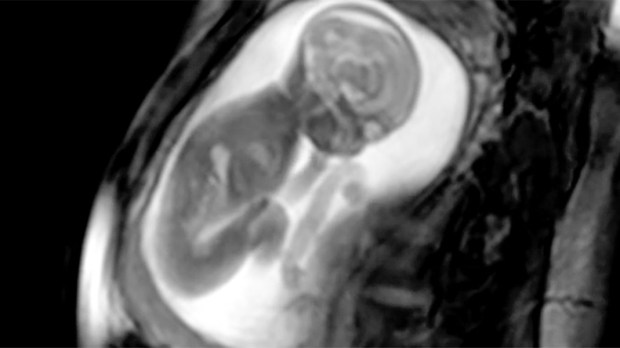
So take a look at these hypnotizing images from an MRI scan of a busy baby showing off its moves in mommy’s belly, proving to us once more just how incredible unborn babies truly are and how they can do a full gymnastic routine in very cramped quarters!

Read more:
She gave her life for her unborn baby — here’s what her husband has to say

Read more:
You can love your unborn baby even if you don’t love pregnancy